A Locally Advanced Papillary Thyroid Carcinoma in a Thyroglossal Duct Cyst Presenting as a Benign Cervical Mass: A Case Report
Ace Joseph C. de la Rosa, Maria Karen A. Capuz
Jul 2021 DOI 10.35460/2546-1621.2018-0077
Introduction
Thyroglossal duct cyst (TGDC) is the most commonly encountered midline and upper cervical mass in the pediatric population with no gender predilection [1-4] and the only true malignancy of the thyroglossal duct is squamous cell carcinoma.[5,6]
Although it is the most common congenital anomaly of the neck and approximately 7% of the general population have this condition [7-9], these patients can also have a rare form of malignancy of this tract, which is papillary carcinoma with cases accounting for only <1%.[1] Current literature on the management of these cases [1,4] is yet to have a consensus in definitive management due to different presentations on the diagnosis as seen in this case.
This case report shows that a Sistrunk procedure alone is also an adequate means for curative treatment.[1]
Case Presentation
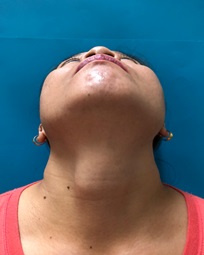
This is a case of a 43 year-old female with a chief complaint of anterior neck mass on clinic visit. History started 10 months prior, when the patient noted a 1x1x1 cm firm, non-tender, anterior neck mass. There were no accompanying symptoms of dyspnea, dysphagia, palpitations, easy fatigability, hoarseness, heat/cold intolerance and no weight loss or gain noted. The patient consulted a private physician where an ultrasound of the neck was requested, the results of which were unrecalled and the patient was lost to follow-up.(Figure 1A)
Interval history revealed asymptomatic progressive increase in mass size. Five months prior, with the increasing mass size, the patient went for another consult with another physician where a biopsy of the said mass was done which showed a colloid cyst. She was advised observation in the meantime; however, the more pronounced size of the neck mass led the patient to consult at the OPD clinic for further management. A thyroid function test was requested which yielded normal result. An ultrasound of the neck showed a superficial complex mass measuring 2.9x2.1x2.9 cm, 2-3 cm superior to the thyroid gland with no malignant features. The findings were consistent with TGDC, hence the patient was advised surgery.
Intraoperative Findings
Intraoperatively the mass was noted to be firm and unusually adherent to the surrounding strap muscles. The hyoid bone was normal looking with the superior part of the mass seemingly invading the inferior border of the hyoid. The hyoid bone was dissected from the surrounding structures and the anterior part removed along with the specimen to complete the Sistrunk procedure. The inferior part of the cyst was noted to be soft with minimal amber colored fluid inside. There was a 3-4 cm of separation of the TGDC from the isthmus of thyroid gland. There were no unusual masses noted within the thyroid gland upon inspection. The surgery was then completed and a Penrose drain was placed.(Figure 2)
Postoperative Findings and Course
One week postoperatively, an ultrasound of the neck was done which revealed surgically removed previous mass at the superior cervical area. The thyroid gland showed homogenous echo textures consistent with a normal thyroid gland. The histopathology report indicated that the mass was consistent with a thyroglossal duct cyst. The superior portion of the mass however, showed papillary thyroid carcinoma with invasion of the surrounding soft tissue with tumor size of 2.4 cm in single greatest diameter. Lympovascular invasion was not identified; however one lymph node was isolated and was positive for papillary thyroid carcinoma. With these findings, total thyroidectomy was offered to the patient to completely eradicate the malignancy. One week postoperatively, ultrasound of the neck was done which showed the previously noted mass at the superior cervical area to have been surgically removed. The thyroid gland exhibited homogenous echo textures consistent with a normal thyroid gland. In light of the histopathologic findings, the patient was however, not keen on undergoing any further surgery, hence, disease surveillance was alternatively advised to the patient through serial thyroid and neck ultrasound monitoring every 6 months.(Figure 1,2)
Follow-up and Outcomes
The patient was monitored with serial ultrasound postoperatively from 1 month, 3 months and 1 year postoperatively with no recurrence of the disease.
Discussion
The physical examination of these patients are usually equivocal and hence, differentiating a malignant from benign TGDC preoperatively is very difficult.[7,9] Most of the TGDC carcinoma cases show benign characteristics with minimal to nil information acquired through ancillary procedures. Physical findings like cervical lymphadenopathy and fixed hard mass should cause alarm for malignancy. In the case of our patient, physical examination from her first visit up until the preoperative assessment showed typical, benign characteristics of a TGDC with no findings suggestive of malignancy. The diagnosis of thyroglossal duct cyst carcinoma (TDCCa) needs a histological demonstration of the duct with associated respiratory epithelium, squamous epithelium or a combination of both.(Figure 3)
This is a rare case without a well-defined management and staging criteria. As such, it has been a cause of debates regarding the optimal management as well as extent of completeness of surgery. Similar cases of TGDCCa have usually been diagnosed postoperatively due to the acellular nature of the cyst contents on fine needle aspiration biopsy.[1,4] In the case of this patient, a Sistrunk procedure was performed based on the preoperative findings consistent for a benign tumor. Intraoperatively however, unexpectedly, a solid, fixed and firm mass adherent to the hyoid bone with infiltration to the surrounding strap muscles was seen. There was note of a 3-4 cm separation of the inferior border of the mass to the thyroid isthmus and thyroid gland.(Figure 2,4,5)
This case had typical benign preoperative clinical findings that conflicted with the suspiciously malignant intraoperative findings causing some challenges in management decisions. The thyroid gland was benign-looking on intraoperative inspection. The distance of the cyst from the gland was significant enough that it was not deemed necessary to remove the thyroid gland at that point in time. It is also worthy to note that on preoperative ultrasonography, the thyroid gland was relatively normal-looking with no heterogeneity on scan, with benign-looking lymph nodes and no thyroid lesions appreciated. Having taken all of this clinical information into consideration, a simple Sistrunk procedure was deemed enough.(Figure 2-5)
Conclusion
Papillary thyroid carcinoma of the TGDC is a rare form of carcinoma from a congenital benign anomaly. Flexibility and clear, open communication with the patient and their relatives are needed in preparation for these kinds or surgery. The management plans should be adequately discussed, especially in cases where there may be features highly suggestive of malignancy yet unproven preoperatively, and a deviation from the original plan for benign lesions may be converted to a more radical surgery as indicated by the intraoperative findings to assure complete eradication of the malignancy.
-
Thompson LDR, Herrera HB, Lau SK. Thyroglossal duct cyst carcinomas: A clinicopathologic series of 22 cases with staging recommendations. Head and Neck Pathology. 2017 Jun;11(2):175–85. DOI: 10.1007/s12105-016-0757-y.
-
Allard RHB. The thyroglossal cyst. Head & Neck [Internet]. 1982 Nov;5(2):134–46. Available from: http://dx.doi.org/10.1002/hed.2890050209
-
Mondin V, Ferlito A, Muzzi E, Silver CE, Fagan JJ, Devaney KO, et al. Thyroglossal duct cyst: Personal experience and literature review. Auris Nasus Larynx [Internet]. 2008 Mar;35(1):11–25. Available from: http://dx.doi.org/10.1016/j.anl.2007.06.001
-
Thompson LDR, Herrera HB, Lau SK. A clinicopathologic series of 685 thyroglossal duct remnant cysts. Head and Neck Pathol. [Internet] 2016 May 9;10(4):465–74. Available from: http://dx.doi.org/10.1007/s12105-016-0724-7
-
Iakovou I, Konstantinidis I, Doumas A, Nikolaidis V, Karatzas N, Efstratiou I. Squamous cell carcinoma in a thyroglossal duct cyst and 99mTc-MIBI findings. Hell J Nucl Med. 2011 Jan-Apr;14(1):62–4.
-
Nuttall FQ. Cystic metastases from papillary adenocarcinoma of the thyroid with comments concerning carcinoma associated with thyroglossal remnants. The American Journal of Surgery [Internet]. 1965 Apr;109(4):500–5. Available from: http://dx.doi.org/10.1016/s0002-9610(65)80185-4
-
Jang DW, Sikora AG, Leytin A. Thyroglossal duct cyst carcinoma: case report and review of the literature. Ear, Nose, & Throat Journal. 2013 Sep;92(9):E12–14.
-
Lira Medina AK, Fernandez Berdeal E, Bernal Cisneros E, Betancourt Galindo R, Frigerio P. Incidental papillary thyroid carcinoma in thyroglossal duct cyst case report. International Journal of Surgery Case Reports. [Internet]. 2016;29:4–7. Available from: http://dx.doi.org/10.1016/j.ijscr.2016.10.021
-
Shah S, Kadakia S, Khorsandi A, Andersen A, Iacob C, Shin E. Squamous cell carcinoma in a thyroglossal duct cyst: A case report with review of the literature. American Journal of Otolaryngology. [Internet] 2015 May;36(3):460–2. Available from: http://dx.doi.org/10.1016/j.amjoto.2015.01.012
Annexure:
 |  |
Fig. 1 (A) preoperative (B) Postoperative
Intraoperative findings:
 |  |  |
Figure 2: (A) Creation of superior and inferior flap exposing the superior border of the hyoid bone. (B) The superior portion of the mass attached to the inferior border of the hyoid bone with a (C) significant distance from the thyroid gland from the inferior border of the cyst.

Figure 3: (A) Microsections of the thyroglossal duct cyst disclose fibro-collagenous tissue partly lined with ciliated to stratified squamous epithelium

(B) On further scanning, clusters of atypical thyrocytes are seen having papillary formation

Figure 4: (A) Thyroglossal duct cyst with anterior segment of the hyoid bone with the cyst on the (B) inferior portion of the cyst. This structure shows invasion of the surrounding soft tissue(C) but not the skeletal muscles.

Figure 5: (A) Scanning micrograph of the identified lymph node on biopsy. (B) The lymph node shows metastasis of atypical thyrocytes.
![]() CC BY:
Open Access Creative Commons Attribution 4.0 International
License, which permits use, sharing, adaptation, distribution and
reproduction in any medium or format, as long as you give
appropriate credit to the original author(s) and the source, provide a link to the Creative Commons license, and indicate if
changes were made. The images or other third party material in
this article are included in the article’s Creative Commons license, unless indicated otherwise in a credit line to the material.
If material is not included in the article’s Creative Commons license and your intended use is not permitted by statutory regulation or exceeds the permitted use, you will need to obtain permission directly from the copyright holder. To view a copy of this
license, visit http://creativecommons.org/licenses/by/4.0/
CC BY:
Open Access Creative Commons Attribution 4.0 International
License, which permits use, sharing, adaptation, distribution and
reproduction in any medium or format, as long as you give
appropriate credit to the original author(s) and the source, provide a link to the Creative Commons license, and indicate if
changes were made. The images or other third party material in
this article are included in the article’s Creative Commons license, unless indicated otherwise in a credit line to the material.
If material is not included in the article’s Creative Commons license and your intended use is not permitted by statutory regulation or exceeds the permitted use, you will need to obtain permission directly from the copyright holder. To view a copy of this
license, visit http://creativecommons.org/licenses/by/4.0/