Effect of Curcumin Supplementation on Rat Skeletal Muscle Morphology and AMPK Levels: Study Protocol for a Randomized Controlled Trial
Maria Grace A. De Guzman, Veatrix Myrtle P. Cruz, Raymond L. Rosales
Apr 2025 DOI 10.35460/2546-1621.2025-0013
Introduction
Sarcopenia is a generalized disorder of the skeletal muscle associated with increased likelihood of adverse outcomes and mortality.[1] Sarcopenia is closely related to aging and characterized by deterioration in skeletal muscle mass and function (defined by muscle strength and physical performance), eventually resulting in reduced physical capability, compromised performance of cardiopulmonary status, risk of falls, presence of disabilities, poor quality of life, high healthcare spending and even mortality.[2]
The reported prevalence rates in the European region vary from as low as 5% to as high as 25%. Epidemiological studies in Asian countries reported that the prevalence rates vary from as low as 5.5% to more than 25%, with male predominance (5.1% to 21.0% in men vs. 4.1% to 16.3% in women).[3] In addition to advanced age, an individual's lifestyle plays a crucial role in the development of sarcopenia. Regular physical activity and balanced nutritional status greatly influence muscle health and strength.
In terms of pathophysiology, skeletal muscle fibers undergo atrophy, especially the type II or fast-twitch fibers, which have low concentrations of mitochondria and are responsible for short, fast bursts of activity.[4] There is also reduced number of motor units and accumulation of fat tissues within the muscle. Different theories for the pathophysiology have been suggested, such as peripheral neurodegeneration, reduction in sensitivity and production of anabolic hormones, dysfunction in the secretion of cytokines and altered inflammatory mechanism.[5]Peripheral neurodegeneration involves neuronal loss, which is a progressive and irreversible process that increases with age. Reduction in motor neurons decreases the available proportion of motor units, resulting in skeletal muscle fiber denervation.[6]
The potential benefits of nutraceuticals and herbal medicines for the prevention and treatment of sarcopenia have been explored in recent years. These natural products are considered less expensive, more accessible and relatively safe (ie, with low toxicity) compared to drugs and high protein dietary approaches being administered to alleviate muscle pain and delay sarcopenia progression, respectively. It is well-documented that oxidative stress and inflammation have important roles in the pathogenesis of muscle damage and pain occurring in sarcopenia. The beneficial effects of these natural products are due to their ability to modulate age-related transcriptional factors in biochemical pathways leading to muscle inflammation.[7]Other beneficial effects include the prevention of muscle damage and atrophy, muscle regeneration and differentiation, and improvement of muscle strength and muscle resistance to fatigue during vigorous exercise.[7]
Among the different natural products with potential therapeutic benefits for sarcopenia prevention and management, curcumin is one of the most studied. Curcuma longa Linn (C. longa) is a rhizome that belongs to the Zingiberaceae (ginger) family. It is a perennial plant cultivated and widely distributed in Asian nations, including the Philippines. Curcumin is well-known for its antioxidant, anti-inflammatory, and anti-aging properties.[7]Curcuma longa has a complete nutritional profile, ie, it contains carbohydrates, proteins, fibers, and fats, as well as vitamins and minerals such as vitamin C, pyridoxine, calcium, potassium, magnesium, and phosphorus. More than 200 phytochemicals have also been found in C. longa.[8] Additionally, curcumin has been shown to improve systemic markers of oxidative stress[9] and gives protective effects to exercise-induced oxidative stress and inflammation, muscle soreness, and muscle recovery in physically active people.[7] Compared to the novel therapeutic agents being explored and studied, curcumin has the advantage of being recognized and utilized worldwide in different forms for a wide array of potential health benefits. It has been used in foods, eg, as curry in India, tea in Japan, and as a preservative and coloring agent even in Western countries like the United States.[10] With the multiple usages of curcumin, its safety has been long established.
Preclinical studies on the effects of curcumin on muscle disorders suggest that the use of curcumin may be a potential solution to prevent or even treat skeletal muscle decline.[8] Reports have shown that curcumin is also beneficial for several conditions that result in skeletal muscle wasting, such as during systemic infection and inflammation. Furthermore, a recent animal study demonstrated improvement in muscle fatigue post-swimming with curcumin, which may be explained by curcumin’s beneficial effect on gut microbiota.[7]Adding curcumin or turmeric extract (TE) to a prescribed diet significantly improved weight loss at 36 weeks in an experimental animal model.[11] The animal study demonstrated that with TE supplementation, there was an improvement in muscle atrophy in both fast-twitch fibers (gastrocnemius and tibialis anterior), which contract over short durations but fatigue rapidly, as well as slow-twitch muscle fibers (soleus), which produce sustained, small movements.
The general objective of this study is to determine the effects of oral curcumin administration on skeletal muscle using an animal model that similarly demonstrates the course of human sarcopenia. The specific objectives are the following: To determine the effect of low-dose and high-dose curcumin supplementation on gross and histologic morphology of rat gastrocnemius muscles; to determine the safety of low-dose and high-dose curcumin supplementation on gross and histologic morphology of rat major organs; and to quantitatively analyze the AMPK of the rat gastrocnemius muscle tissue.
Methods
The study will last for 35 days (five weeks). An acclimation period of seven days will be allotted to allow animals enough time to stabilize in a new environment and promote both animal welfare and reproducible experimental results. The experiment proper will last for 28 days (four weeks). Purpose-bred 11- to 12-week-old female Sprague Dawley (SD) rats (N = 32) will be used in this study (Figure 1A). These animals have been chosen because they are specifically bred and raised in controlled environments for biomedical research purposes. SD rats are being extensively used in animal models of human diseases and conditions such as diabetes, obesity, cancer, cardiovascular, renal diseases[12],and sarcopenia.[13,14] The sample size calculation was based on the assumption that each subgroup will include at least four animals; if one subject does not survive, there will be at least three remaining. The females have been selected because of the following reasons: (i) compared to males, females do not show more temperature or activity variance;[15] (ii) females are more stable in behavior even if they have an estrous cycle (Caruso, 2023). The animals will be sourced from the Animal Pharmacology Unit of Unilab, Inc., Medical and Regulatory Affairs, Biological Sciences Department.
Laboratory Animal Care Procedure
The study will be conducted at the Laboratory Animal Facility (LAF) of the Animal Pharmacology unit of the Biological Sciences Department (AP-BSD) of Unilab, Inc., Medical and Regulatory Affairs because of the following reasons: (i) It is equipped with a well-maintained microenvironment, ie, the immediate physical environment surrounding the animal such as cage and feeding equipment (Figure 1C and 1D). (ii) There are individual ventilated cages (IVC) where animals are protected by High Efficiency-Particulate Air (HEPA)-filters that protect them from all micro-organisms. (iii) The Animal Pharmacology (AP) unit ensures that the animal's health status is maintained, following the animal welfare guidelines and meeting scientific requirements: regular health monitoring and microbial monitoring. (iv) There is an established policy for entry and exit in animal facilities (Figure 1E and F). The AP unit personnel wear personal protective equipment (PPE) when inside the animal laboratory premises (Figure 1B). The AP unit personnel are immunized with anti-rabies and anti-tetanus vaccines. (v) The unit has a total of four animal caretakers going on duty eight hours daily, including holidays, Saturdays, and Sundays. (vi) The AP-BSD LAF has been granted a certificate of registration (or License to Operate) by the Philippine Department of Agriculture, Bureau of Animal Industry (BAI)[16] in accordance with the provision of the Republic Act No. 3639.

Figure 1. (A) Sprague-Dawley rat; (B) AP-BSD personnel wearing personal protective equipment; (C) drinking water bottles; (D) Polycarbonate (Techniplast, 480 x 265 x 210 mm) cages with stainless steel cage covers; (E) Entrance to the Laboratory Animal Facility; (F) Designated area for changing to laboratory coats and other protective clothing (Source: AP-BSD, Unilab Research Compound).
The study animals (SD rats) will be kept individually in sterilized polycarbonate cages (480 x 265 x 210 mm) with stainless steel covers having provisions for holding pellet feed and drinking water bottles fitted with stainless steel sipper tubes. The cage range meets and exceeds current international rules and guidelines. The animals will be maintained as per standard protocol throughout the study.[17] The animals will be provided with appropriate feeding and housed in individual cages within temperature and humidity ranges appropriate for their species, to which they can adapt with minimal stress and physiologic alteration. The primary purpose of ventilation is to provide appropriate air quality and adequate oxygen supply. The rats are nocturnal animals, and photoreceptors in their eyes are adapted to dim lighting conditions between 1 and 40 lux. The environmental condition will include the following: Room temperature: 22˚C (±3˚C); Relative Humidity: 50%-70%; Ventilation: 10 to 15 air changes/hour; Lighting: 12 hours light/12 hours dark cycle. Autoclaved rice hulls will be used as beddings, with a depth of at least 2 cm. The animal diet and feeding will include the following: Sarimanok® (sourced from Univet Nutrition and Animal Healthcare Company, UNAHCO, Inc.) rodent pellets will be given as feeds. Purified water (sourced from the Reverse Osmosis Water Filtration System, LAF of Unilab, Inc. Medical and Regulatory Affairs) will be given ad libitum.
The animal cages will be washed thoroughly twice a week with ordinary detergent soap and solution with an antimicrobial agent in a designated area of the LAF. During cleaning, the animals will be transferred to temporary clean cages with exact specifications as the animals’ permanent cages. All clean cages will be dried with clean paper towels. Water bottles and food containers will be washed with antimicrobial dishwashing liquid, rinsed thoroughly, and wiped dry. Waste materials and used beddings will be placed in a yellow trash bag in a designated trash bin for disposal.
Animal models utilized in sarcopenia studies (Figure 2) include the use of aged laboratory animals (with expected age-related skeletal muscle atrophy and denervation), hind limb unloading, denervation, and immobilization.[13,18,19] The changes occurring in skeletal muscles of aged rats are almost similar to the changes in sarcopenia among humans. However, since the natural aging process will require longer period of time to build, it will not be feasible to utilize it as an ideal model for this study. Furthermore, laboratory animals require prolonged housing and feeding, and thus, higher maintenance cost, and their availability primarily limits their use as natural sarcopenia models. Limiting physical activities, unloading (to eliminate weight), and immobilization (to reduce muscle mass) are common strategies involved in low-activity for sarcopenia models.[18] The tail suspension (TS), a simpler model introduced by Nemoto and Goyagi(2021), is a low-activity method utilizing hind limb unloading, which involves decreased hind limb function by suspending the animal’s tail for at least two weeks (Figure 3).

Figure 2. Rodent model of muscular atrophy for sarcopenia study (Source: Baek, et al., 2020, p. 98, Fig. 1).


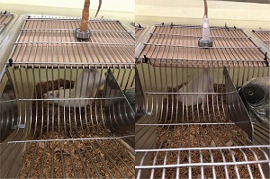
Figure 3. Tail suspension. - (A) and (B) Rats were suspended by their tail. Free access to food and water is allowed without the hind limbs touching the ground. (Source: AP-BSD, Unilab Research Compound).
General description of animal manipulation methods
-
Handling and Restraining: For safe and effective handling, the handler must have comprehensive knowledge of the animal’s anatomy, physiology, and responsive behavior. Steps should be taken to allow rats to become familiar with the researcher who will be handling them so as to reduce the stress of handling. This should include the process of “gentling,” whereby rats are allowed to explore their handler and are gently stroked and held.[20,21]Restraint may significantly cause stress on laboratory animals, including rodents. The stress caused by daily handling and feeding gavage insertion may be reduced when the act is done repeatedly; this is called the habituation process.[22]

Figure 4. Handling and restraining techniques: (A) Using his/her dominant hand, the researcher/research assistant will grasp the rat’s tail and pull it gently. (B) The researcher/research assistant will put his/her non-dominant hand over the rat’s back. (C) The rat will be grasped around the thorax, with the researcher/research assistant’s index finger over the rat’s shoulder and his/her middle, ring and small fingers wrapping around the rat’s thorax, forming a “V” (light green color). Sources: Manzoor, et al., 2013, Figure 4 for A and B; Cruz, LC lecture proceeding, 2021) -
Hind Limb Unloading (TS): The bodies of laboratory animals will be suspended by the tail using improvised non-harmful materials to prevent the hind limbs from touching the ground (Figure 3A), for two to four weeks, with free access to food and water (Figure 3B). A cloth medical tape will be used to anchor the rat’s tail to the upper portion of the cage. A cushioning (foam) will be added before the clip is fitted in order to avoid direct contact between the tail and clip. The thickness and fit of cushioning will be adjusted accordingly to prevent possible injury, such as tail ischemia. The behavior of animals will be observed regularly during the first few days of adjustment to the hind limb unloading method.
-
Oral Administration: Oral substances are administered to animals utilizing gavage tubes made up of stainless steel to prevent animals from biting them. The gavage tubes or needles are also called feeding needles or feeding tubes (Figures 5A and 5B). All gavage needles have a ball- or pear-shaped smooth rounded tip (Figure 4A, tip encircled) to prevent injury to the esophagus and other tissues.[23] The size of gavage needles to be used will be based on the animal's body weight.[23] Each rat will be weighed individually to determine the appropriate gavage needle size. The maximum amount that can be administrated orally is 20 ml/kg for rodents.[22] The length of gavage needle is measured from the tip of the rat’s nose to its xiphoid process (corresponding to the bottom part of the sternum bone). This procedure will show how far the needle will be inserted into the esophagus. Oral gavage procedure should only be performed by trained personnel (ie, the research assistant). The rat will be properly restrained prior to oral administration of the test substance. The trained research assistant will hold the rat’s body near the thoracic region with its lower body supported and caution should be observed to prevent restricting the rat’s respiration. The rat’s head should be gently positioned in an extended manner to create a straight line from the neck and esophagus. During actual feeding, the end tip of the gavage needle will be inserted into the rat’s mouth and allowed to slide along the roof of the rat’s oral cavity towards the left side with gravity alone (ie, the personnel will not force the needle into the rat’s esophagus). The gavage needle should easily pass into the esophagus in one direction only (clockwise or counter-clockwise) and without any resistance. Resistance will be encountered when feeding tube is accidentally inserted into the trachea (the windpipe), which may cause the animal's sudden death. If a wrong insertion happens, the tube should be withdrawn immediately, and a second insertion trial should be done. Only two insertion attempts should be made to avoid stress to the animal. The test drug (diluted with a specified diluent such as distilled water to become a solution) will be slowly administered by a syringe attached to the end of the needle once the gavage needle is appropriately in place. There should be no undue rotation of the gavage needle to prevent injury to the esophagus. After oral administration, the needle will be gently removed following the same angle as insertion, and the animal will be returned to its cage.[23]


Figure 5. Reusable stainless feeding gavage needles or tubes (https:// gavageneedle.com/products/reusable-oral-gavage-needle )
Administration of Interventions/Treatments
The study animals will be randomly divided into four groups (Figure 6), the control (CON, n = 8), the vehicle (VEH, n = 8), the low-dose curcumin (CUR, n = 8) and the high-dose CUR (n = 8) utilizing the randomization function in Microsoft Excel. The curcumin has been sourced from Herbanext Laboratories Inc., Negros South Road, Bago City, Negros Occidental, Philippines. Propylene glycol (PG) will be used as the vehicle. It is a water-soluble fluid considered by the US Food and Drug Administration as a Generally Recognized As Safe (GRAS) additive, ie, a safe substance in foods, drugs, and cosmetics.[24] The oral format of PG is quickly and widely absorbed, with a rapid distribution to tissues in proportion to body water. Elimination from the body is through metabolic clearance and renal excretion. PG has a low degree of toxicity in both humans and animals.[25] In humans, there was no lethal oral dose reported. However, for laboratory rats, the minimal lethal oral dose is reported to be 20.9 grams/kilogram.[24] In vehicle control, the apparently safe substance is used alone and administered similarly to the rest of the treatment arms. When compared with the untreated control (or placebo), the vehicle control will determine whether vehicle administration alone causes any effects.[26,27]Only the two CUR groups will receive the test substance (a local curcumin formulation) in a single dose per day using an oral gavage feeding technique by trained personnel. The control group will be given an equal volume of distilled water (without curcumin). The curcumin doses will be computed per body weight. The low dose is 100 mg/kg/day, which was approximated based on previous animal studies utilizing 100 mg/kg/day and 170 mg/kg/day, respectively,[28,29] while the high dose is 1000 mg/kg/day, based on the previous animal study.[30]

Figure 6: Allocation of interventions (test substances)
Animal Examination Procedures
Periodic monitoring of each animal will be done during the first 24 hours and daily thereafter, until the end of the study period. To facilitate monitoring, a camera will be utilized to do daily observations and examinations, which will include deaths (mortality), general state, external appearance, behavior, clinical symptoms, and changes in body weight. The individual weights of all animals will be obtained before initiation of the test substance and at least weekly thereafter. The changes in weight measurements will be calculated and recorded. The following parameters will be observed daily: general state and appearance (including changes in skin and fur, eyes and mucous membranes); respiratory, circulatory, autonomic, and central nervous systems; somatomotor activity and behavior pattern. The animal examination will be performed based on the protocol stated in the Pharmacology-Toxicology section of “A Guidebook to Plant Screening: Phytochemical and Biological” as provided by the Research Center for the Natural and Applied Sciences of the University of Santo Tomas (UST-RCNAS).[31] The following signs and symptoms will also be observed: tremors, convulsions, salivation, diarrhea, lethargy, sleep, and coma. All observations will be captured and recorded through video and photographs, and findings reported. All surviving animals will be weighed and humanely killed at the completion of the study. When humane endpoints such as loss of 20% body weight, cachexia, dehydration, respiratory distress, or weakness (eg, inability to reach food and water) are observed, the animal/s should be euthanized or treated to relieve unnecessary pain or distress.
Euthanasia, Post-Mortem Examination and Animal Disposal
-
Euthanasia: It is the act of inducing humane death in an animal by a method that induces rapid loss of consciousness and death with a minimum of pain, discomfort, or distress.[32]Trained personnel should perform the method, which should be reliable and irreversible and with minimal restraint. The method must comply with the most recent guidelines on euthanasia published by the American Veterinary Medical Association (AVMA). Carbon dioxide (CO2) inhalation is the most common method of euthanasia used for rodents, such as mice, rats, guinea pigs, gerbils, and hamsters. The only acceptable source of CO2 is compressed gas because this permits gas inflow to the induction chamber (covered cage) to be controlled.[32] The proper operating procedure should be carefully followed because the anesthetic effects of CO2 are reversible. Animals that are prematurely removed from the chamber may recover.[32]To confirm the laboratory animal's death, close observation will be conducted. If there are indications that the animal is still alive, cervical dislocation will be performed.[33]
-
Gross Necropsy: The organs will be harvested by a licensed veterinarian. Materials required for pathological examination include a dissection board, forceps, scissors, labels for containers, and fixative and collection containers (eg, tubes, cups) should be prepared. The procedure of organ collection and examination will be in accordance with the Revised Guides for Organ Sampling and Trimming in Rats and Mice.[34] All major organs in the thoraco-abdominal area (heart, lungs, gastrointestinal, kidneys, reproductive) will be examined for abnormalities like color changes and size differences. The thoracic and abdominal cavities will be examined for any presence of fluid/s.
-
Animal Disposal: The death of animals will be ensured by doing a second physical examination. The carcasses will be placed in two leak-proof bags. All bags with animal carcasses must be labeled with the following information: IACUC Number, method used to ensure death, date, and initials of the person disposing of the carcass. Once sealed, the bags will be brought to the designated place of the Unilab, Inc. Biological Sciences Department for proper disposal.
Dissection and Examination of Skeletal Muscle
-
Skeletal Muscle Dissection: The femoral and tibial bones will be dissected, and gastrocnemius muscles on both sides will be excised and weighed. Thereafter, muscle tissues will be subjected to rapid freezing in acetone-dry ice and sliced into 10 μm-thick sections for Hematoxylin and eosin (H&E), Nicotinamide Adenine Dinucleotide tetrazolium reductase (NADH), and Gomori trichrome staining.
-
Histologic Examination: Together with the examination of muscle architecture (for degeneration, necrosis, nuclear orientation, central nucleation, fibrosis, and singular/group atrophy), computation of atrophy factors will also be performed. The presence of connective tissue, fat tissue, and the number of atrophic muscle cells will also be determined.[35]
AMPK Determination
Accurate quantitative detection of the rat total AMP-Activated Protein Kinase (AMPK) will be performed in the gastrocnemius muscle tissue utilizing the enzyme-linked immunosorbent assay (ELISA) kit. The kit, which is based on the Double antibody-sandwich ELISA detection method, will be utilized for in vitro quantitative determination of AMPK concentrations in serum, plasma, cell culture supernatant, and other biological samples such as muscle tissue. The RAT AMPK ELISA Kit (Catalog number ER0730, Unit Product ID Q09137) will be sourced from Fine Biotech (FineTest).[36] The method will follow the standard protocol described in the product information.
AMPK is an intracellular energy sensor inhibiting ATP consumption and also stimulating ATP production under energy-depleted conditions [37] and is considered a key regulator of skeletal muscle metabolism.[38] Exercise has been considered the most powerful physiological activator of AMPK,[39] and activation of its α2 unit was demonstrated during intensive physical exercise.[40] Animal studies on various natural products have demonstrated activation of AMPK even when given alone, without any form of exercise.[35] Both curcumin alone and exercise (with or without curcumin) significantly increased AMPK phosphorylation.[41]
Statistical Analysis
Animal data (baseline, week 2, and week 4) will be presented in tabular form as the mean ± standard deviation. Outcome variables will be analyzed using one-way ANOVA, with significance set a priori at p<0.05. The variables to be analyzed include whole body weight, gastrocnemius muscle weight, gastrocnemius muscle size at specified time points, and atrophy factors. For the quantification of AMPK using ELISA, statistics will be calculated using GraphPad Prism. One-way or two-way ANOVA will be performed, and multiple comparison analysis will be done to evaluate significance between groups. Gross and histological skeletal muscle findings will be presented in a descriptive form. All deaths, whether immediate, delayed, or humane kills, will be incorporated for the purpose of maximum likelihood analysis. The confidence interval calculations will be done using the computer program package.
Ethics and Dissemination
-
Ethics Approval: The study will comply with the Philippine animal welfare laws. The study protocol has been reviewed by the Institutional Animal Care Use Committee of the University of Santo Tomas (UST IACUC), Manila, Philippines.
-
Dissemination: The final data will be shared with the public regardless of study outcomes. Results will be presented at relevant conferences and submitted to an appropriate journal following data analysis and trial closure.
Contributors: The corresponding (principal) author designed the study, and will gather, analyze, interpret data, and complete the manuscript. All other authors will contribute to data collection and critically review the manuscript.
Competing Interests: RLR is part of the editorial board of JMUST. VPC is employed as a veterinarian of Unilab, Inc., Medical and Regulatory Affairs Division.
- Cruz-Jentoft AJ, Bahat G, Bauer J, Boirie Y, Bruyère O, Cederholm T, et al. Sarcopenia: revised European consensus on definition and diagnosis. Age Ageing [Internet]. 2019;48(4):601. Available from: http://dx.doi.org/10.1093/ageing/afz046
- Chen L-K, Liu L-K, Woo J, Assantachai P, Auyeung T-W, Bahyah KS, et al. Sarcopenia in Asia: consensus report of the Asian Working Group for Sarcopenia. J Am Med Dir Assoc [Internet]. 2014;15(2):95–101. Available from: http://dx.doi.org/10.1016/j.jamda.2013.11.025
- Chen L-K, Woo J, Assantachai P, Auyeung T-W, Chou M-Y, Iijima K, et al. Asian Working Group for sarcopenia: 2019 consensus update on sarcopenia diagnosis and treatment. J Am Med Dir Assoc [Internet]. 2020;21(3):300-307.e2. Available from: http://dx.doi.org/10.1016/j.jamda.2019.12.012
- Talbot J, Maves L. Skeletal muscle fiber type: using insights from muscle developmental biology to dissect targets for susceptibility and resistance to muscle disease. Wiley Interdiscip Rev Dev Biol [Internet]. 2016;5(4):518–34. Available from: http://dx.doi.org/10.1002/wdev.230
- Kim TN, Choi KM. Sarcopenia: definition, epidemiology, and pathophysiology. J Bone Metab [Internet]. 2013;20(1):1–10. Available from: http://dx.doi.org/10.11005/jbm.2013.20.1.1
- Pascual-Fernández J, Fernández-Montero A, Córdova-Martínez A, Pastor D, Martínez-Rodríguez A, Roche E. Sarcopenia: Molecular pathways and potential targets for intervention. Int J Mol Sci [Internet]. 2020;21(22):8844. Available from: http://dx.doi.org/10.3390/ijms21228844
- Bagherniya M, Mahdavi A, Shokri-Mashhadi N, Banach M, Von Haehling S, Johnston TP, et al. The beneficial therapeutic effects of plant-derived natural products for the treatment of sarcopenia. J Cachexia Sarcopenia Muscle [Internet]. 2022;13(6):2772–90. Available from: http://dx.doi.org/10.1002/jcsm.13057
- Vargas-Mendoza N, Madrigal-Santillán E, Álvarez-González I, Madrigal-Bujaidar E, Anguiano-Robledo L, Aguilar-Faisal JL, et al. Phytochemicals in skeletal muscle health: Effects of curcumin (from Curcuma longa Linn) and sulforaphane (from Brassicaceae) on muscle function, recovery and therapy of muscle atrophy. Plants [Internet]. 2022;11(19):2517. Available from: http://dx.doi.org/10.3390/plants11192517
- Sahebkar A, Serban M-C, Ursoniu S, Banach M. Effect of curcuminoids on oxidative stress: A systematic review and meta-analysis of randomized controlled trials. J Funct Foods [Internet]. 2015;18:898–909. Available from: http://dx.doi.org/10.1016/j.jff.2015.01.005
- Hewlings SJ, Kalman DS. Curcumin: A review of its effects on human health. Foods [Internet]. 2017;6(10). Available from: http://dx.doi.org/10.3390/foods6100092
- Lyu W, Kousaka M, Jia H, Kato H. Effects of turmeric extract on age-related skeletal muscle atrophy in senescence-accelerated mice. Life (Basel) [Internet]. 2023;13(4). Available from: http://dx.doi.org/10.3390/life13040941
- Brower M, Grace M, Kotz CM, Koya V. Comparative analysis of growth characteristics of Sprague Dawley rats obtained from different sources. Lab Anim Res [Internet]. 2015;31(4):166–73. Available from: http://dx.doi.org/10.5625/lar.2015.31.4.166
- Nemoto A, Goyagi T. Tail suspension is useful as a sarcopenia model in rats. Lab Anim Res [Internet]. 2021;37(1):7. Available from: http://dx.doi.org/10.1186/s42826-020-00083-9
- Shu H, Huang Y, Zhang W, Ling L, Hua Y, Xiong Z. An integrated study of hormone-related sarcopenia for modeling and comparative transcriptome in rats. Front Endocrinol (Lausanne) [Internet]. 2023;14:1073587. Available from: http://dx.doi.org/10.3389/fendo.2023.1073587
- Smarr B, Kriegsfeld LJ. Female mice exhibit less overall variance, with a higher proportion of structured variance, than males at multiple timescales of continuous body temperature and locomotive activity records. Biol Sex Differ [Internet]. 2022;13(1):41. Available from: http://dx.doi.org/10.1186/s13293-022-00451-1
- Republic Act No. 3639: An Act Creating the Bureau of Animal Industry, Defining its Powers and Functions [Internet]. Philippine Veterinary Medical Association. [cited 2025]. Available from: https://www.pvma.com.ph/republic-acts#tab-1-5
- Trivedi MK, Branton A, Trivedi D, Jana S. Prevention of aging and improvement of longevity and life-span in D-galactose induced aging rats after treatment with the biofield energy per se and biofield treated proprietary test formulation. J Ageing Res Healthc [Internet]. 2020;3(1):48–57. Available from: http://dx.doi.org/10.14302/issn.2474-7785.jarh-20-3425
- Baek K-W, Jung Y-K, Kim J-S, Park JS, Hah Y-S, Kim S-J, et al. Rodent model of muscular atrophy for sarcopenia study. J Bone Metab [Internet]. 2020;27(2):97–110. Available from: http://dx.doi.org/10.11005/jbm.2020.27.2.97
- Palus S, Springer JI, Doehner W, von Haehling S, Anker M, Anker SD, et al. Models of sarcopenia: Short review. Int J Cardiol [Internet]. 2017;238:19–21. Available from: http://dx.doi.org/10.1016/j.ijcard.2017.03.152
- Manzoor M, Raza S. Proficient handling and restraint of the laboratory animal rat (rattus norvegicus) facilitate essential biochemical and molecular level studies in biomedical sciences. IOSR Journal of Pharmacy and Biological Sciences. 2013;6:21–33.
- ARRP Guideline 20: Guidelines for the Housing of Rats in Scientific Institutions [Internet]. Orange NSW, AU: Animal Welfare Branch, NSW Department of Primary Industries; 2008 [cited 2025]. Available from: https://www.animalethics.org.au/__data/assets/pdf_file/0014/222512/housing-rats-scientific-institutions.pdf
- Turner PV, Brabb T, Pekow C, Vasbinder MA. Administration of substances to laboratory animals: routes of administration and factors to consider. J Am Assoc Lab Anim Sci. 2011;50(5):600–13.
- Standard Operating Procedure #10. Standard Operating Procedures for Oral Gavage in Mice and Rats [Internet]. Washington State University Institutional Animal Care and Use Committee (WSU IACUC). 2021 [cited 2025]. Available from: https://iacuc.wsu.edu/documents/2016/06/wsu_sop_10.pdf/
- HEALTH EFFECTS. In: Toxicological Profile for Propylene Glycol [Internet]. Atlanta (GA): Agency for Toxic Substances and Disease Registry (US); 1997 [cited 2025]. p.7–65. Available from: https://www.ncbi.nlm.nih.gov/books/NBK598030/
- McMartin K. Propylene Glycol. In: Encyclopedia of Toxicology [Internet]. 3rd ed. Elsevier; 2014. p.1113–6. Available from: http://dx.doi.org/10.1016/b978-0-12-386454-3.01029-0
- Robinson NB, Krieger K, Khan FM, Huffman W, Chang M, Naik A, et al. The current state of animal models in research: A review. Int J Surg [Internet]. 2019;72:9–13. Available from: http://dx.doi.org/10.1016/j.ijsu.2019.10.015
- Johnson PD, Besselsen DG. Practical aspects of experimental design in animal research. ILAR J [Internet]. 2002;43(4):202–6. Available from: http://dx.doi.org/10.1093/ilar.43.4.202
- Sahin K, Pala R, Tuzcu M, Ozdemir O, Orhan C, Sahin N, et al. Curcumin prevents muscle damage by regulating NF-κB and Nrf2 pathways and improves performance: an in vivo model. J Inflamm Res [Internet]. 2016;9:147–54. Available from: http://dx.doi.org/10.2147/JIR.S110873
- Rinkunaite I, Simoliunas E, Alksne M, Dapkute D, Bukelskiene V. Anti-inflammatory effect of different curcumin preparations on adjuvant-induced arthritis in rats. BMC Complement Med Ther [Internet]. 2021;21(1):39. Available from: http://dx.doi.org/10.1186/s12906-021-03207-3
- Hocking AJ, Elliot D, Hua J, Klebe S. Administering fixed oral doses of curcumin to rats through voluntary consumption. J Am Assoc Lab Anim Sci [Internet]. 2018;57(5):508–12. Available from: http://dx.doi.org/10.30802/AALAS-JAALAS-17-000143
- Guevara BQ. A Guidebook to Plant Screening; Phytochemical and Biological. Manila, Philippines: Research Center for the Natural and Applied Sciences, University of Santo Tomas; 2004.
- Animal welfare regulations require that the Attending Veterinarian (AV) shall provide guidance to the Principal Investigator (PI) and staff regarding euthanasia, and that the Institutional Animal Care and Use Committee (IACUC) must approve the method of euthanasia to be utilized for research animals. [Internet]. University of Nebraska Omaha (UNOMAHA) Institutional Animal Care and Use Committee. 2021 [cited 2025]. Available from: https://www.unomaha.edu/office-of-research-and-creative-activity/research-compliance/iacuc/iacuc-policies/policy-for-euthanasia-and-carcass-disposal.pdf
- Sivula CP, Suckow MA. Euthanasia. In: Management of Animal Care and Use Programs in Research, Education, and Testing [Internet]. 2nd ed. Boca Raton : Taylor & Francis, 2018. CRC Press; 2017. p.827–40. Available from: http://dx.doi.org/10.1201/9781315152189-35
- Ruehl-Fehlert C, Kittel B, Morawietz G, Deslex P, Keenan C, Mahrt CR, et al. Revised guides for organ sampling and trimming in rats and mice--part 1. Exp Toxicol Pathol [Internet]. 2003;55(2–3):91–106. Available from: http://dx.doi.org/10.1078/0940-2993-00311
- Çinar İ, Bozoğlan M, Aytekin K, Esenyel D, Esenyel CZ. The histopathological effects of sleep disorders on striated muscle in rats. Saudi Med J [Internet]. 2023;44(4):355–62. Available from: http://dx.doi.org/10.15537/smj.2023.44.4.20220714
- Rat AMPK (phosphorylated adenosine monophosphate activated protein kinase) ELISA kit [Internet]. FineTest ELISA Kit | FineTest Antibody | FineTest®. Wuhan Fine Biotech Co., Ltd.; 2019 [cited 2025]. Available from: https://www.fn-test.com/product/er0730/
- Kim T, Davis J, Zhang AJ, He X, Mathews ST. Curcumin activates AMPK and suppresses gluconeogenic gene expression in hepatoma cells. Biochem Biophys Res Commun [Internet]. 2009;388(2):377–82. Available from: http://dx.doi.org/10.1016/j.bbrc.2009.08.018
- Thomson DM. The role of AMPK in the regulation of skeletal muscle size, hypertrophy, and regeneration. Int J Mol Sci [Internet]. 2018;19(10). Available from: http://dx.doi.org/10.3390/ijms19103125
- Richter EA, Ruderman NB. AMPK and the biochemistry of exercise: implications for human health and disease. Biochem J [Internet]. 2009;418(2):261–75. Available from: http://dx.doi.org/10.1042/BJ20082055
- Wojtaszewski JF, Nielsen P, Hansen BF, Richter EA, Kiens B. Isoform-specific and exercise intensity-dependent activation of 5’-AMP-activated protein kinase in human skeletal muscle. J Physiol [Internet]. 2000;528 Pt 1:221–6. Available from: http://dx.doi.org/10.1111/j.1469-7793.2000.t01-1-00221.x
- Hamidie RDR, Shibaguchi T, Yamada T, Koma R, Ishizawa R, Saito Y, et al. Curcumin induces mitochondrial biogenesis by increasing cyclic AMP levels via phosphodiesterase 4A inhibition in skeletal muscle. Br J Nutr [Internet]. 2021;126(11):1642–50. Available from: http://dx.doi.org/10.1017/S0007114521000490
![]() Open Access This article is licensed under a Creative Commons Attribution-NonCommercial-ShareAlike 4.0
International License, which permits use, share — copy and redistribute the material in any medium or format,
adapt — remix, transform, and build upon the material, as long as you give appropriate credit,
provide a link to the license, and indicate if changes were made. You may do so in any reasonable manner,
but not in any way that suggests the licensor endorses you or your use. You may not use the material for
commercial purposes. If you remix, transform, or build upon the material, you must distribute your
contributions under the same license as the original. You may not apply legal terms or technological
measures that legally restrict others from doing anything the license permits. The images or other
third party material in this article are included in the article’s Creative Commons license, unless indicated
otherwise in a credit line to the material. If material is not included in the article’s Creative Commons
license and your intended use is not permitted by statutory regulation or exceeds the permitted use,
you will need to obtain permission directly from the copyright holder. To view a copy of this license,
visit https://creativecommons.org/licenses/by-nc-sa/4.0/.
Open Access This article is licensed under a Creative Commons Attribution-NonCommercial-ShareAlike 4.0
International License, which permits use, share — copy and redistribute the material in any medium or format,
adapt — remix, transform, and build upon the material, as long as you give appropriate credit,
provide a link to the license, and indicate if changes were made. You may do so in any reasonable manner,
but not in any way that suggests the licensor endorses you or your use. You may not use the material for
commercial purposes. If you remix, transform, or build upon the material, you must distribute your
contributions under the same license as the original. You may not apply legal terms or technological
measures that legally restrict others from doing anything the license permits. The images or other
third party material in this article are included in the article’s Creative Commons license, unless indicated
otherwise in a credit line to the material. If material is not included in the article’s Creative Commons
license and your intended use is not permitted by statutory regulation or exceeds the permitted use,
you will need to obtain permission directly from the copyright holder. To view a copy of this license,
visit https://creativecommons.org/licenses/by-nc-sa/4.0/.